Who we are

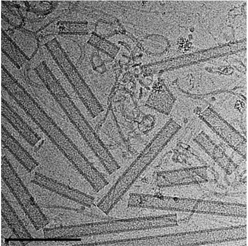
We tailor high resolution imaging techniques to study the mechanism of ESCRT driven membrane fission during cytokinesis of mammalian cells.

Since the invention of the first light microscope in 1670, history tell us that new developments in microscopy induce a leap in our understanding of cell biology.
Our moto is to advance state-of-the-art microscopy techniques including quantitative live cell imaging and super resolution (SR) microscopy in order to resolve how molecular machines organize in cells to execute their biological function.

Specifically, we focus on understanding how the ESCRT complex orchestrate in cells to cut membranes. By constantly seeking for new information that will take us closer to understanding the mechanism of ESCRTs in their native environment, we aim to advance both biology and microscopy.

Because the tools that we develop are driven by our experimental needs we are confident that they will be broadly used by cell biologists to study other complex cellular processes. Read more

Lab news

Congratulation to Inna for the recent publication in Cell Reports

Though it wasn’t easy, it showed that hard work and a collaborative lab effort eventually pay off.  In her work Inna showed that: 3D STROM can be used to resolve the structure of the ESCRT machine in cytokinesis. ESCRT-III organizes into narrowing cortical spirals to drive abscission. Abscission involves polymerization and depolymerization of the ESCRT-III …

Research

Cellular function of ESCRTs

At the end of mammalian cell division, the two nascent daughter cells are connected by a narrow membrane tube called the intercellular bridge. Cutting of the intercellular bridge, termed abscission, is driven by the ESCRT complex. The spatiotemporal characteristics of abscission are ideal for high-end microscopy techniques. To elucidate the mechanism of ESCRTs in physiological context we therefore apply high-resolution microscopy techniques to visualize ESCRTs in mammalian cell abscission. To obtain an inclusive understanding of ESCRT function in abscission, we visualize abscission in both mammalian tissue culture cells and in a developmental system (zebrafish embryogenesis). By combining information obtained from a variety of microscopy tools including live cell imaging, X-ray tomography and Super resolution microscopy we generate mechanistic models for ESCRT-mediated membrane abscission.

Read more

Advancing microscopy

High-resolution fluorescence imaging, combined with labeling techniques available today have yet been able to provide the spatiotemporal resolution needed to document cellular processes at physiological conditions. Substituting the use of fluorescent protein tags (such as GFP) typically used in live cell applications with fluorescent dyes will improve the currently advanced imaging techniques considerably. The superb photophysical characteristics of Fl-dyes and their small size sizes make them superior probes for quantitative high-resolution fluorescence imaging, particularly in the study of macromolecular assemblies.

Read more

Our tool box

We invest a great effort in developing ways to resolve the structural organization of ESCRT complex in abscission at sufficient spatiotemporal resolution. Over the past few years we established a microscopy-based toolbox to reach our goal.

Read more